The new Gateway MRI Center for Biomolecular Imaging at Gateway University Research Park in Greensboro, North Carolina consists of interdisciplinary scientists with expertise in structural and functional imaging.
Housed in a state-of-the-art building at the Joint School for Nanoscience and Nanoengineering (JSNN), our imaging scientists collaborate across disciplinary fields to exploit emerging frontiers in neural engineering and develop new approaches for determining anatomical and functional connectivity in neural circuits. These frontiers include recording, imaging, analyses and manipulating activity in networks and defined ensembles of neurons.
Our Gateway MRI Center experimental work is tightly coupled with the theoretical and computational efforts carried out by scientists within JSNN and the University of North Carolina at Greensboro, along with regional, national, and global partners.
Fostering vibrant investigations and research efforts across disciplinary boundaries, the Gateway MRI Center ultimately will comprise interdisciplinary research groups that combine state-of-the art imaging with emerging analytics to explore the bio-inspired mechanisms of thought, feeling, movement and perception.
SCANNER TECHNOLOGY
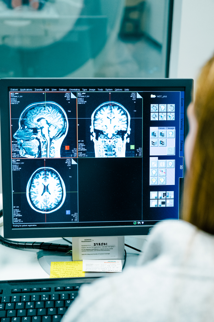
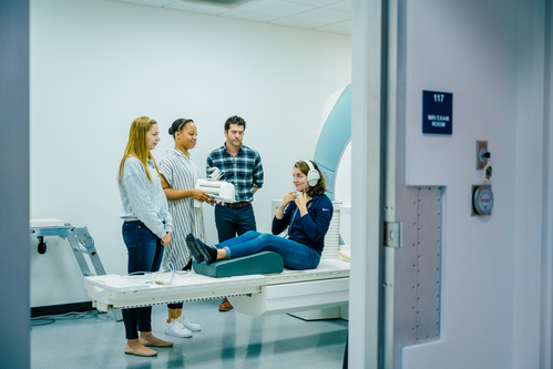
The facility is equipped with a Siemens Magnetom Tim Trio 3 Tesla MRI scanner with a 60cm center inner bore diameter and a 50 cm spherical diameter imaging volume. The scanner has a variety of RF coils to accommodate the broad imaging needs of faculty.
COMPUTING INFRASTRUCTURE AND PROCESSING SOFTWARE
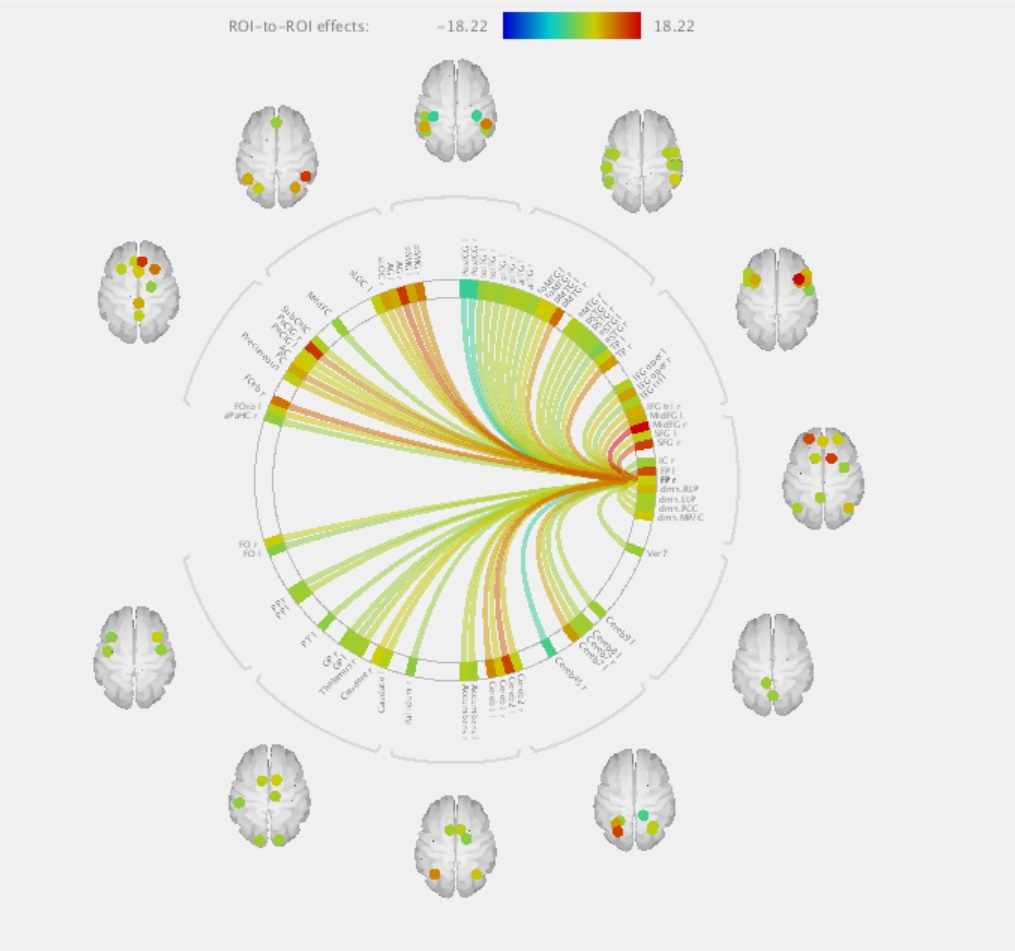
All images acquired on the Siemens scanner are stored offsite behind the UNCG firewall on a secure PACS server. Offline network storage with automatic backups is available to all UNCG investigators. This offline storage is mounted to a server with 16 cores (Xeon X5560) operating at 2.8 GHz with 8 Mbytes of internal cache and 64 GBytes total memory. The server is dedicated for scientific computing and is available for processing and analysis of all MRI data. Software packages presently installed on this server include Slicer 3D, Matlab, FSL, Free Surfer, SPM8, CONN Toolbox, GraphVar, Diffusion Toolkit, BrainNet Viewer and ITK-SNAP. More software options are available. Please contact the Gateway MRI Center Directors for information on how to upload potential new software.